


Search
Search
Saksonov Stanislav; Teslenko Olexandr; and Dmytrulina Nikolietta
Show Description +
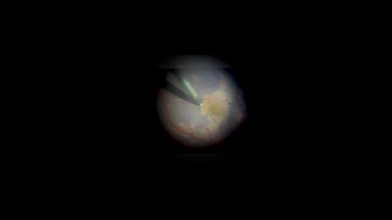
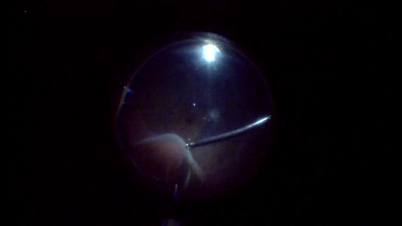
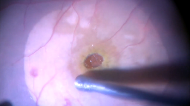
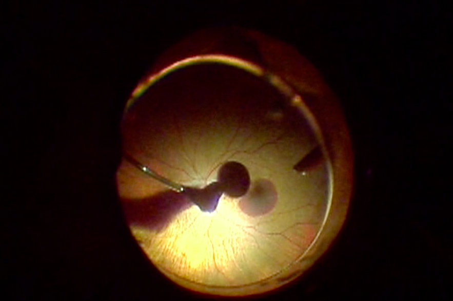
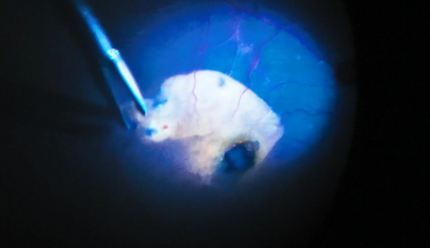
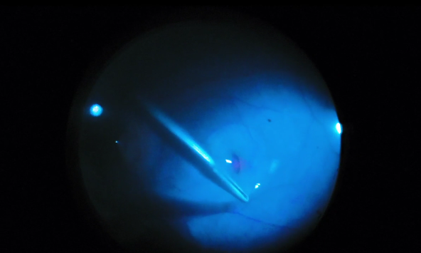
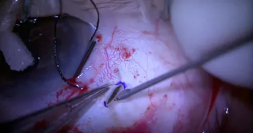
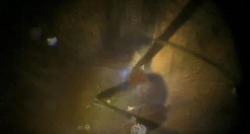
The speakers demonstrate surgical management of a 101-year-old woman who presented with a sudden deterioration of vision in her left eye for the past 3 days. Fundus examination of the left eye revealed a Hollenhorst plaque located at the inferior branch of the central retinal artery. Despite approximately 72 hours having passed since the onset of symptoms, the cholesterol embolus remained clearly visible and was located directly at the origin of the retinal arterial branch from the optic disc. This created an opportunity to mechanically target the cause of the occlusion; therefore, the speakers performed transluminal Nd:YAG laser embolysis to fragment the embolus directly within the vascular lumen and thereby attempt to restore blood flow to the distal retinal circulation.
Posted: 10/15/2026
Saksonov Stanislav; Teslenko Olexandr; and Dmytrulina Nikolietta
The speakers demonstrate surgical management of a 101-year-old woman who presented with a sudden deterioration of vision in her left eye for the past 3 days. Fundus examination of the left eye revealed a Hollenhorst plaque located at the inferior branch of the central retinal artery. Despite approximately 72 hours having passed since the onset of symptoms, the cholesterol embolus remained clearly visible and was located directly at the origin of the retinal arterial branch from the optic disc. This created an opportunity to mechanically target the cause of the occlusion; therefore, the speakers performed transluminal Nd:YAG laser embolysis to fragment the embolus directly within the vascular lumen and thereby attempt to restore blood flow to the distal retinal circulation.
Posted: 10/15/2026





Please log in to leave a comment.