


Search
Search

How data and personal experience are evolving the treatment of geographic atrophy.

Geeta Lalwani, MD; Murtaza Adam, MD; and Carl Danzig, MD
Show Description +
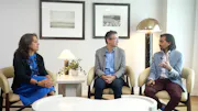
What imaging biomarkers signal a higher risk of rapid progression in geographic atrophy, and how might AI eventually help track treatment effect in real time? Moderator Geeta Lalwani, MD, and panelists Murtaza Adam, MD, and Carl Danzig, MD, review specific multimodal imaging approaches at the point of diagnosis, the field's growing comfort with earlier intervention with complement inhibitors, and the technology gaps still standing in the way of measuring GA progression on an individual level.
Posted: 7/01/2026
Geeta Lalwani, MD; Murtaza Adam, MD; and Carl Danzig, MD
What imaging biomarkers signal a higher risk of rapid progression in geographic atrophy, and how might AI eventually help track treatment effect in real time? Moderator Geeta Lalwani, MD, and panelists Murtaza Adam, MD, and Carl Danzig, MD, review specific multimodal imaging approaches at the point of diagnosis, the field's growing comfort with earlier intervention with complement inhibitors, and the technology gaps still standing in the way of measuring GA progression on an individual level.
Posted: 7/01/2026
Read Transcript
Geeta Lalwani, MD:
Thanks for sticking with us. I'm going to turn the discussion now to biomarkers and how you use them in practice and how you see them playing out in the future. So Carl, talk to me when you first meet a patient, we've discussed this, that you use multimodal imaging. What do you look at first?
Carl Danzig, MD:
I always look at the OCT first, probably because it's easier to look at. I can scroll through it nicely. I use the Heidelberg Spectalis and I'm looking for that hypertransmission defect to really look at is there CROR, complete atrophy of the outer retina and RPE. I also do autofluorescent imaging and I'm looking for multifocal extrafoveal lesions. I'm looking if there's a hyperautofluorescence and I'm looking for bilaterality. Those are really the big four I'm looking at. And that's what I tell patients about that as a risk for more rapid progression. We never really know how fast someone's going to progress. And on top of that, we can't measure how fast someone progress on or off treatment, but we talk about this is a risk factor for you to potentially lose your vision faster than had you not had these risk factors.
Murtaza Adam, MD:
Patients fall on a bell curve. And so you can have a patient present with a unifocal circular lesion that's 1500 plus microns away from the fovea and there's no hypoautofluorescence or hyperautofluorescence surrounding a lesion. That's a patient that's low risk. But if they have a larger lesion that has irregular borders, it's more like a puzzle piece as opposed to something that's purely circular, circularity actually is a protective factor against growth of GA. So that's another risk factor that you look at. And then to your point about bilaterality, if the other eye has geographic atrophy that's foveal involving and larger and their fellow eye that sees better has multifocal GA, I always highlight to the patient that there are differences between these eyes, but they're headed down the same path. And so those features really, really attune me to counsel patients more aggressively.
Geeta Lalwani, MD:
Sure. So once you start treatment, do you do anything different in terms of imaging?
Carl Danzig, MD:
Not really. I'll still get an OCT at every single visit because I want to make sure that a patient does develop wet AMD while on anti-complement therapy. And I will continue getting the autofluorescent photos every six months indefinitely. And I'm hoping that there will be some technology in the future that will allow us to measure progression.
Murtaza Adam, MD:
Yeah, you make such a good point. I think in a way the cart came before the horse. We have these drugs to treat GA. We're excited to use them, but we lack the ability to demonstrate that we're reducing the progression to the fovea and the actual area of GA in the real world. We just don't have the technology to say, this is the GA area progression that we've seen over time and here's how it's slowing down. So our patients are reassured that we're making an effect. A lot of it is based on our qualitative imaging that we take on a monthly or every month basis along with our Q6 month to Q year autofluorescence images. So it's a challenge that we face.
Carl Danzig, MD:
And some of our patients are super highly educated, as you mentioned before, and they want to know the difference between wet AMD and dry AMD treatment or GA treatment. And I explained that in wet AMD treatment, the therapy came along when a new diagnostic test came along also. So you had OCT that showed efficacy of therapy. So a new therapy that you can measure to efficacy quickly, non-invasively at every visit. We're not there yet with GA.
Murtaza Adam, MD:
We're close. We just need interoperability between platforms. So as an example, we have a platform that we use to measure GA area. We primarily use it to pre-screen for clinical trials, but if they could just incorporate that metric into our OCT readouts, how great would that be? We could show patients that we're actually making a dference as opposed to saying, well, we believe the clinical trial data, which we do, but really you want to know what the individual patient's doing.
Geeta Lalwani, MD:
Sure. It seems like it lends itself really well to an AI powered progression assessment for each of these patients telling us what we should be doing with patients who's going to be higher risk of faster progression beyond the biomarkers we discussed with. So we can actually have true data, I think, to decide who would benefit the most from these treatments for sure.
Carl Danzig, MD:
The other thing, Geeta, is that if we get these patients sooner, we can do these images upstream. And then we can truly find out what their rate of progression is. If they have a little extrafoveal lesion that's far away, if it progresses and how it changes with treatment. And then also if we get these patients more upstream, some practices are offering photobiomodulation for intermediate AMD. So that is actually driving some referrals sooner to my practice, but still just not enough.
Geeta Lalwani, MD:
I do think that retina specialist on average are getting comfortable treating GA earlier than when we first did a few years ago when it was first released. What is your own personal experience with that?
Murtaza Adam, MD:
That's totally true. I mean, I think starting with a novel molecule in the real world, although you're reassured by the safety and the clinical trial data that we've seen, you feel more comfortable when a patient's sort of already started to lose vision when you're starting a new therapy. But you're 100% right. As the years have gone on, my comfort of starting treatment in early patients that I think have significant risk of vision loss in the next few years, especially if they're younger patients with high functionality, my threshold's really low to recommend treatment.
Geeta Lalwani, MD:
Sure. I think all the data that has come out, especially regarding the rate of progression of GA two and a half years till it reaches a fovea has been a little bit shocking to some of us.
Murtaza Adam, MD:
Truly humbling to know that we sort of ignored that statistic.
Geeta Lalwani, MD:
Yeah. And we're using FAF and we're watching it progress right in front of our eyes. So I think that it's a good term for patients.




![[2019] Amniotic Membrane Placement for Repair of Recurrent Macular Hole](https://d33x7gwyb2b5z6.cloudfront.net/eyetube/thumbs/gldaw.jpg?auto=compress,format&w=504)
Please log in to leave a comment.