


Search
Search
Show Description +
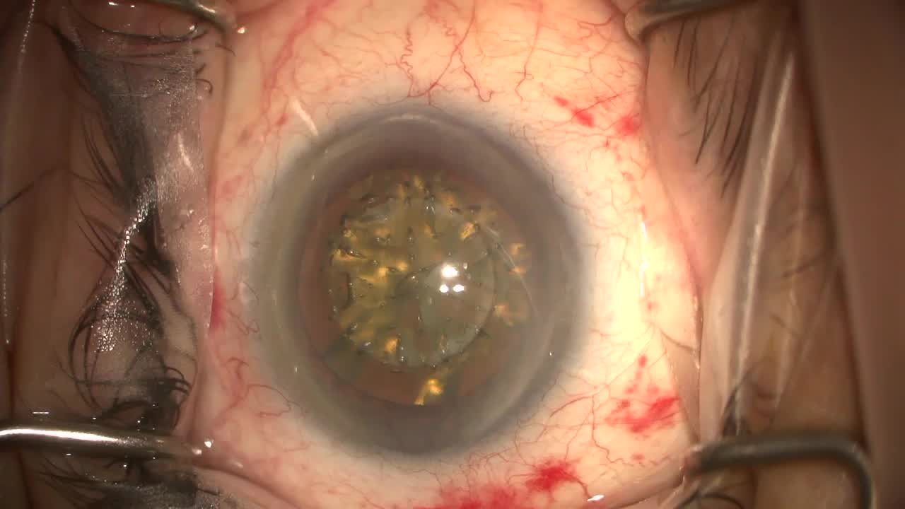
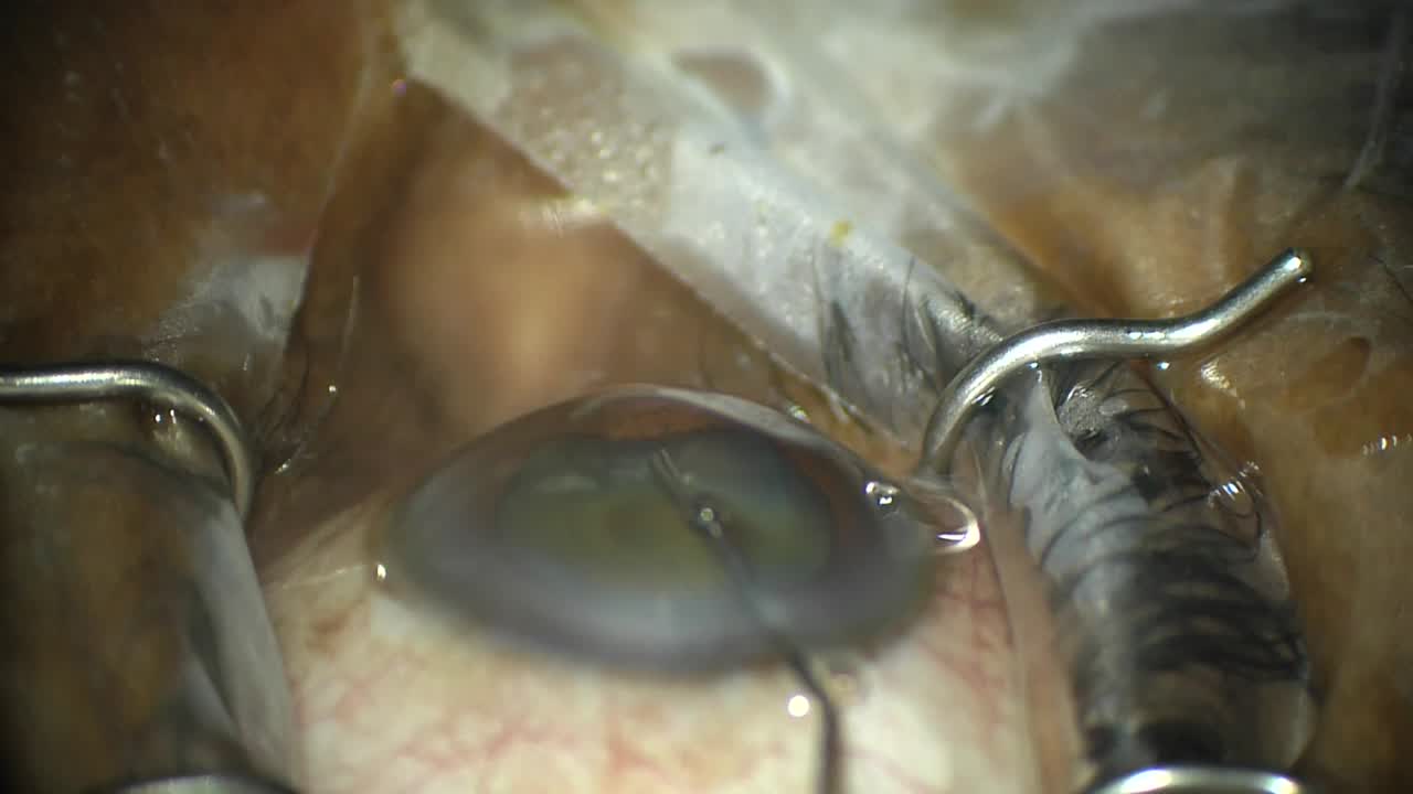
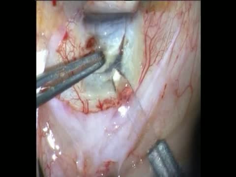
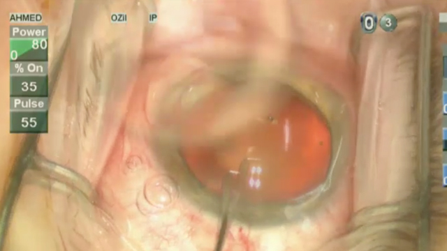
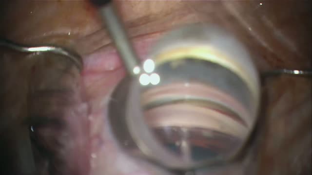
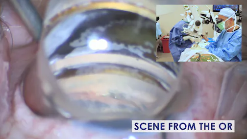
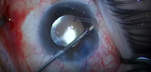
Steven Vold, MD, demonstrates the benefits of transscleral cyclophotocoagulation. He uses the G-Probe (Iridex) handpiece and indents the conjunctiva and centers the fiber optic tip 1.2 mm from the corneoscleral limbus, oriented parallel to the visual axis. While the design of the G-Probe is a significant aid in the delivery of laser energy to the correct location, Dr. Vold also believes the use of transillumination is essential. He has a technician shine a fiber optic light source from the side, tangential to the lens, to light the globe so he can see exactly where the ciliary processes are.
Posted: 10/18/2012
Steven Vold, MD, demonstrates the benefits of transscleral cyclophotocoagulation. He uses the G-Probe (Iridex) handpiece and indents the conjunctiva and centers the fiber optic tip 1.2 mm from the corneoscleral limbus, oriented parallel to the visual axis. While the design of the G-Probe is a significant aid in the delivery of laser energy to the correct location, Dr. Vold also believes the use of transillumination is essential. He has a technician shine a fiber optic light source from the side, tangential to the lens, to light the globe so he can see exactly where the ciliary processes are.
Posted: 10/18/2012


Please log in to leave a comment.