


Search
Search
Show Description +
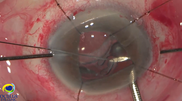
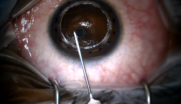
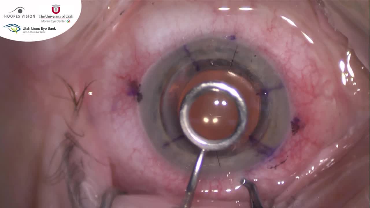
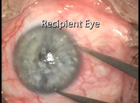
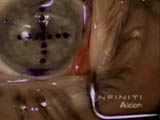
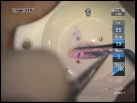
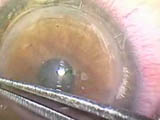
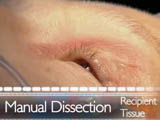
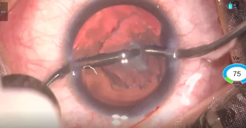
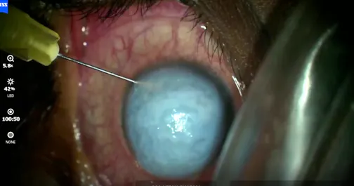
Alex W. Cohen, MD, PhD, and Ben Kunz, MD, perform a DMEK using Iowa Lions Eye Bank prepared tissue. The tissue is punched at 8 mm and marked with an S-stamp to help with orientation. We use a 2.4 mm incision and a B+L Mi60L lens inserter to inject the tissue.
Posted: 2/03/2016
Alex W. Cohen, MD, PhD, and Ben Kunz, MD, perform a DMEK using Iowa Lions Eye Bank prepared tissue. The tissue is punched at 8 mm and marked with an S-stamp to help with orientation. We use a 2.4 mm incision and a B+L Mi60L lens inserter to inject the tissue.
Posted: 2/03/2016



Please log in to leave a comment.