


Search
Search
Show Description +
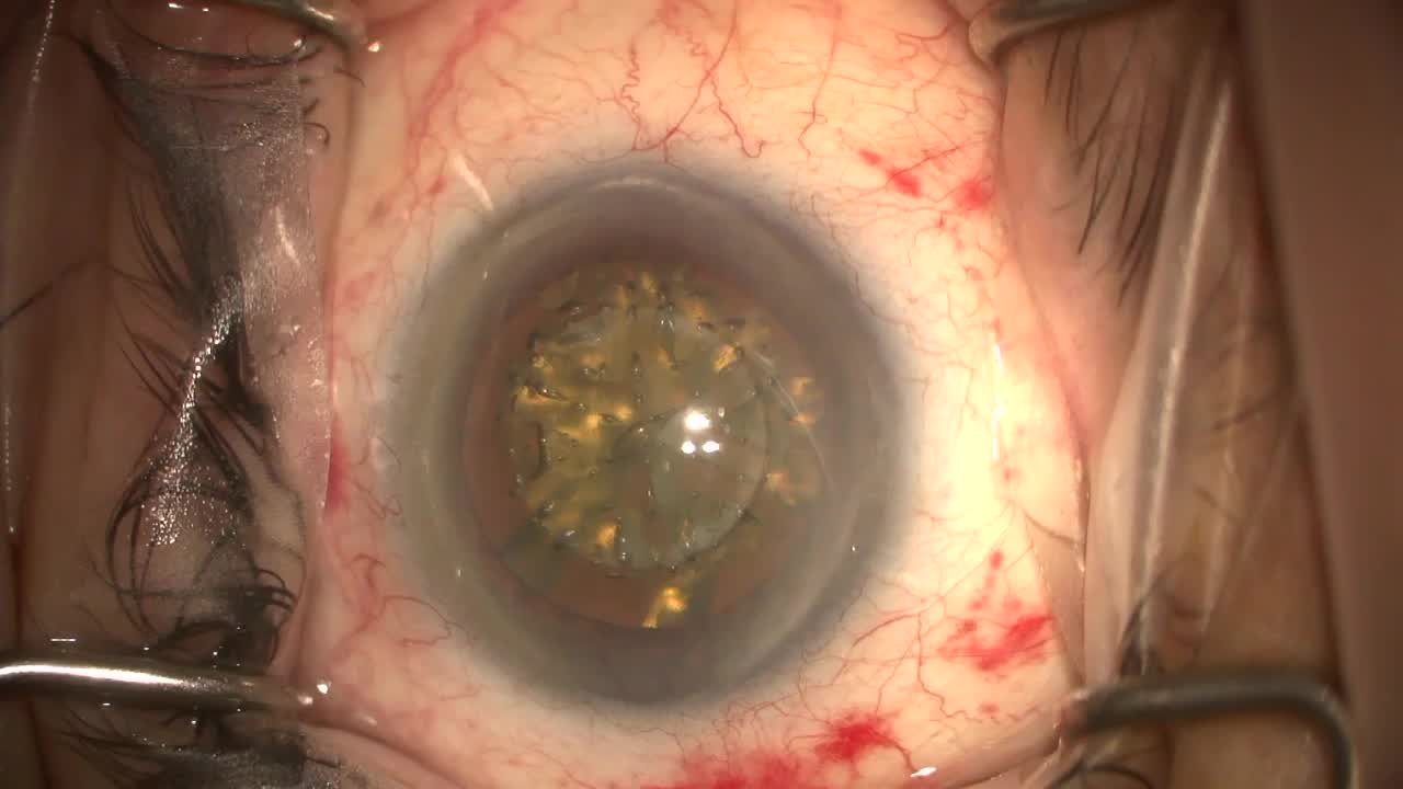
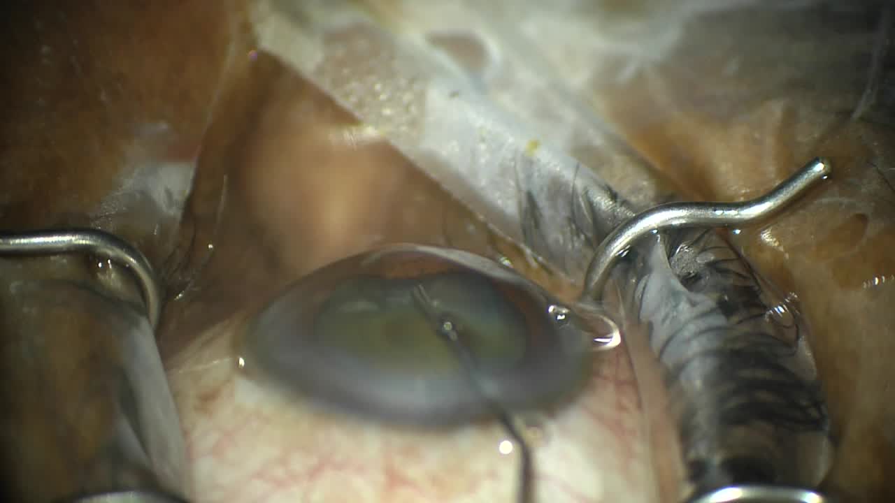
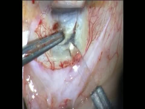
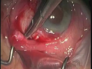
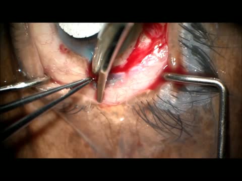
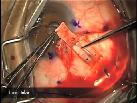
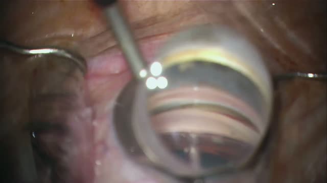
Ahmed Elwehidy, MD, demonstrates a combined procedure in which he performs a circumferential suture trabeculotomy and phacoemsulsification. First, a superior limbal-based conjunctival flap is created, then a superficial rectangular scleral flap is fashioned and dissected toward the limbus. A deep scleral flap is created toward the limbus underneath the superficial flap exposing the scleral spur and deroofing Schlemm canal. A 5-0 polypropylene suture tip is cauterized and inserted into the canal using microsurgical forceps and advanced through the whole circumference of the canal, creating a 360-degree trabeculotomy.
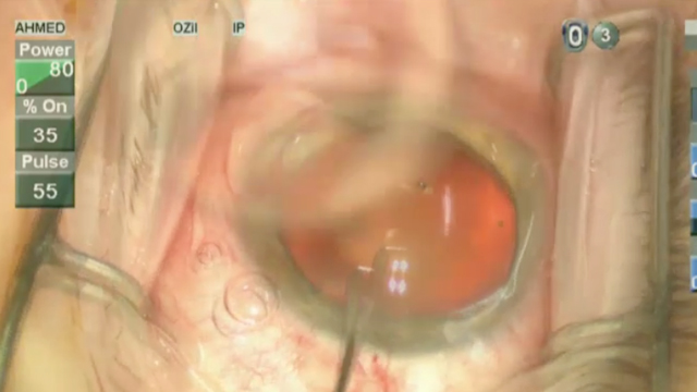
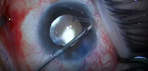
Then, phacoemulsification is started by making a clear corneal incision with a 3.2-mm keratome along with two side port Incisions. Capsulorhexis of the desired size is performed, and the nucleus is emulsified and aspirated through an ultrasonic phaco probe inserted through the main wound. Following this, the remaining cortical matter is removed from the capsular bag, and a foldable IOL is implanted.
Posted: 9/06/2023
Ahmed Elwehidy, MD, demonstrates a combined procedure in which he performs a circumferential suture trabeculotomy and phacoemsulsification. First, a superior limbal-based conjunctival flap is created, then a superficial rectangular scleral flap is fashioned and dissected toward the limbus. A deep scleral flap is created toward the limbus underneath the superficial flap exposing the scleral spur and deroofing Schlemm canal. A 5-0 polypropylene suture tip is cauterized and inserted into the canal using microsurgical forceps and advanced through the whole circumference of the canal, creating a 360-degree trabeculotomy.
Then, phacoemulsification is started by making a clear corneal incision with a 3.2-mm keratome along with two side port Incisions. Capsulorhexis of the desired size is performed, and the nucleus is emulsified and aspirated through an ultrasonic phaco probe inserted through the main wound. Following this, the remaining cortical matter is removed from the capsular bag, and a foldable IOL is implanted.
Posted: 9/06/2023



_1790078971.png?auto=compress,format&w=504)
Please log in to leave a comment.