Search
Search
Enrico Borrelli, MD, PhD, FEBO
Show Description +
Posted: 1/26/2024
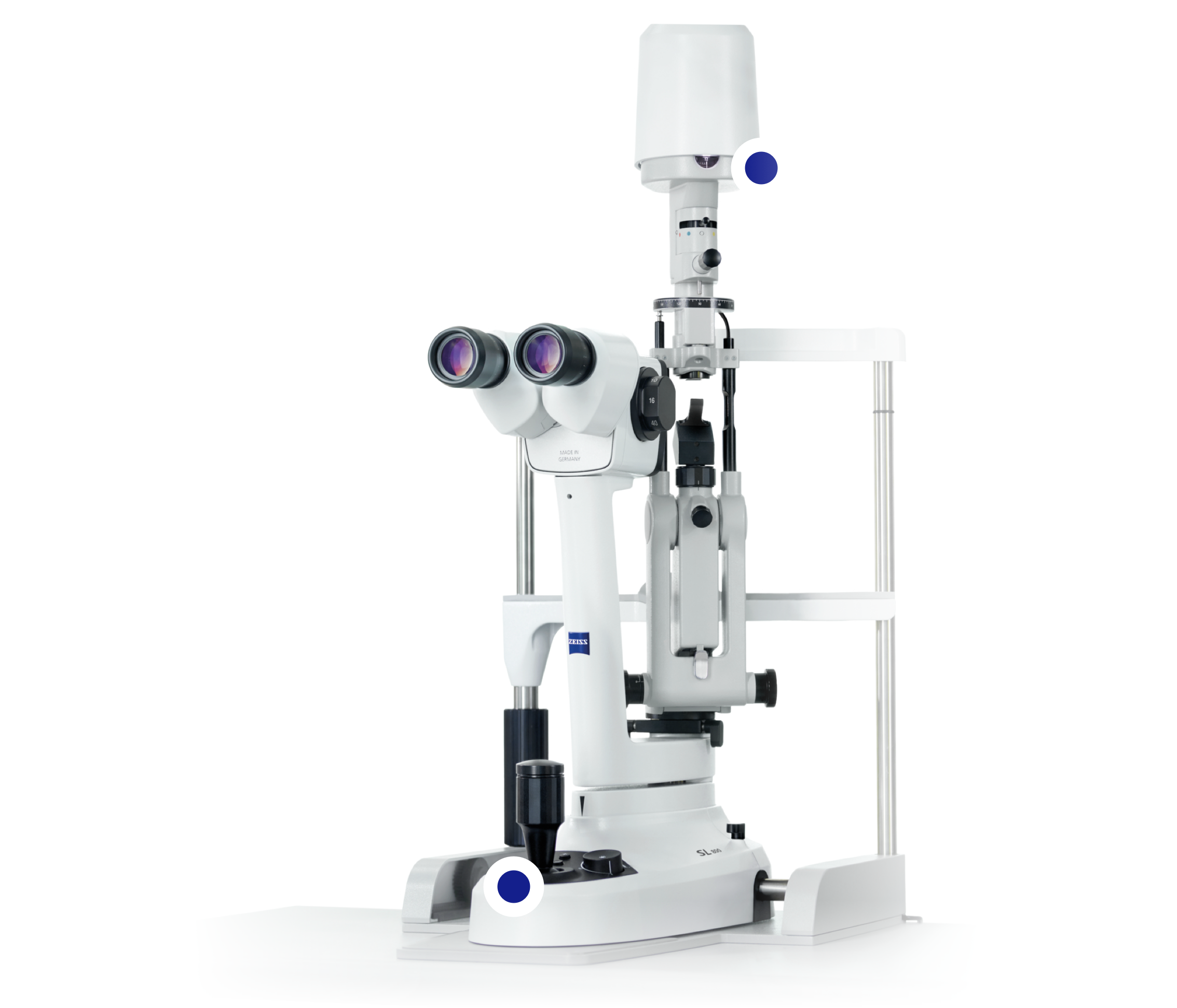
ZEISS Retina Workflow
AMD Management with ZEISS Retina Workflow
ZEISS Retina Workflow solutions for Diabetic Patients

How Imaging Helps Clinics Integrate New Therapies in GA
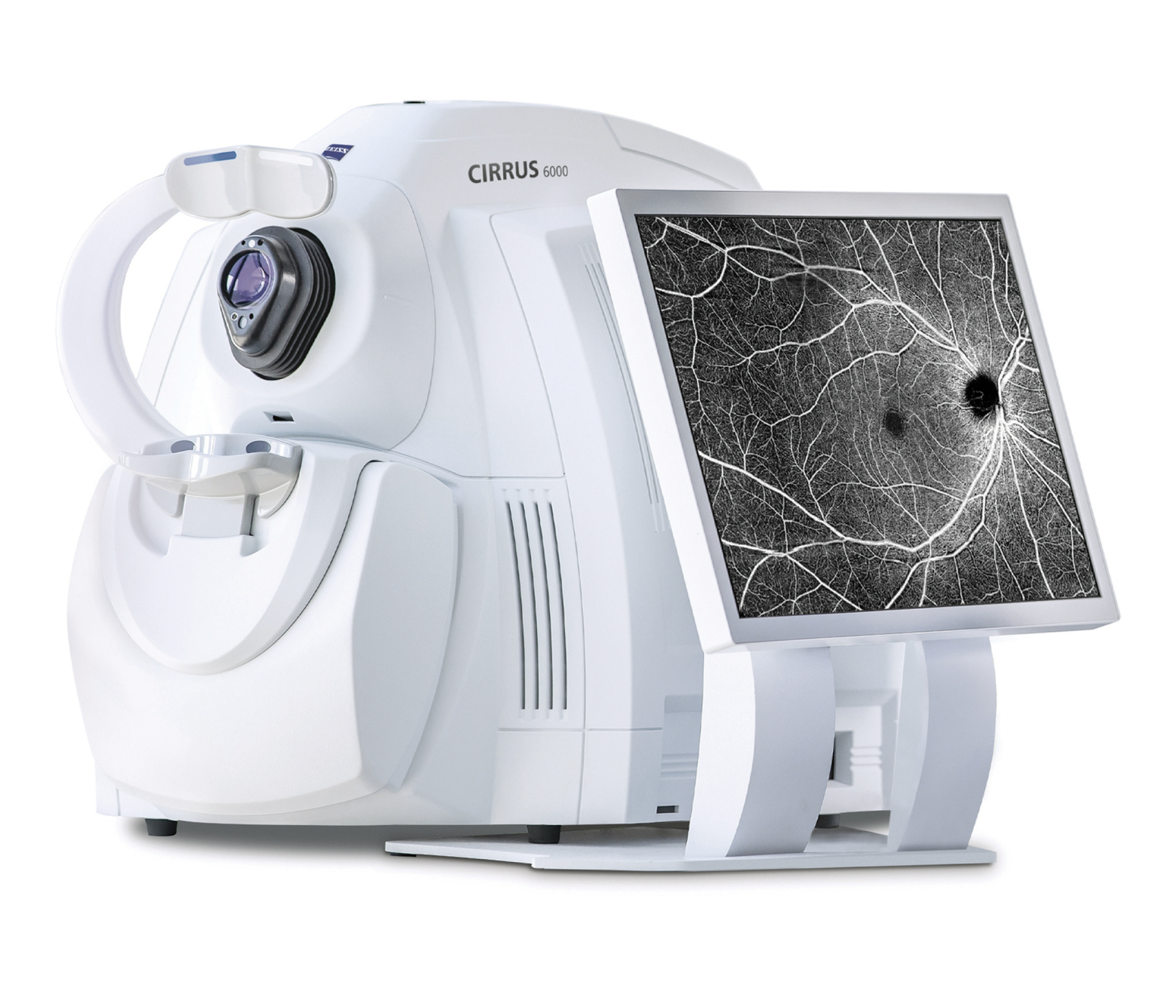
Imaging Geographic Atrophy: OCT, FAF, NIR, and OCTA
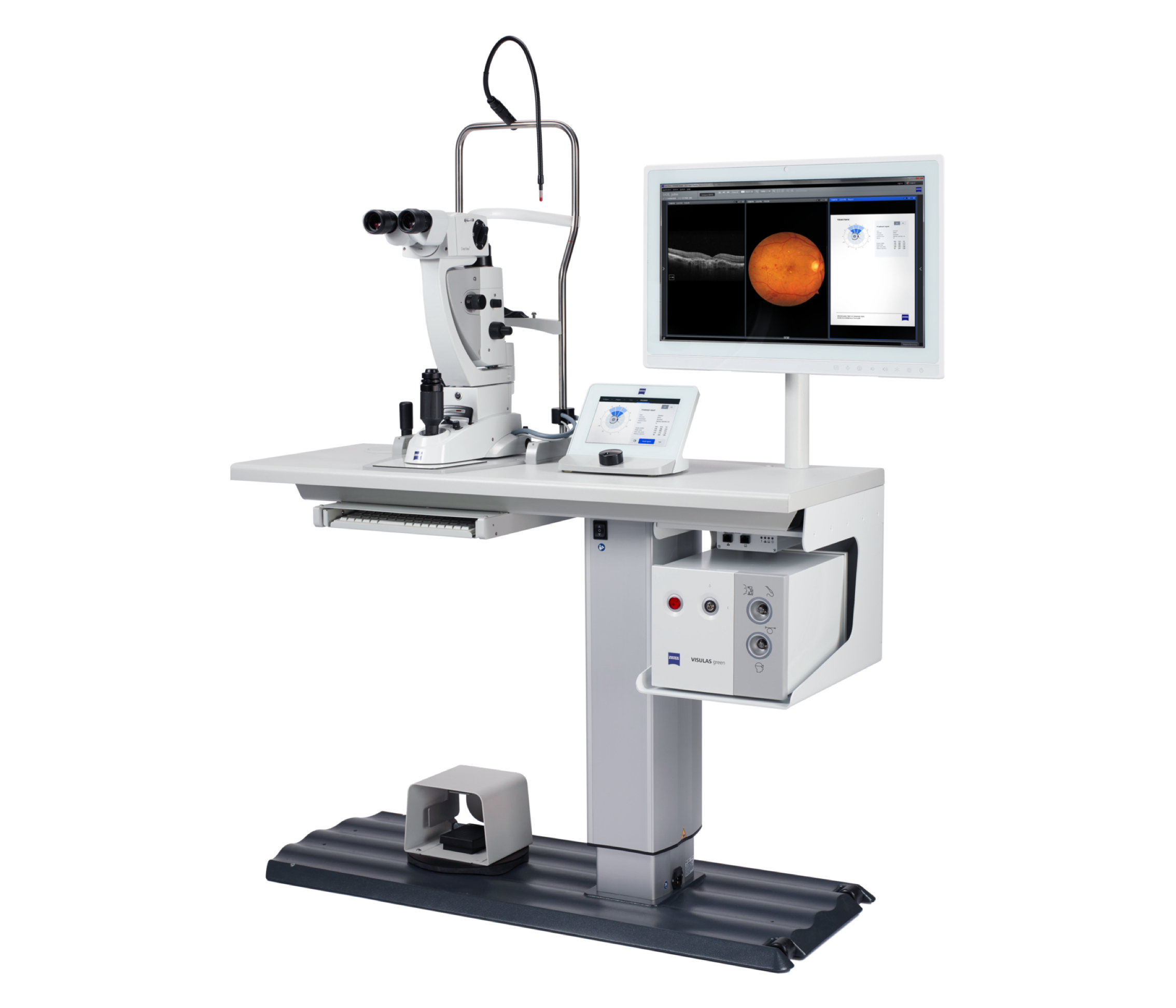
Zeiss Retina Workflow
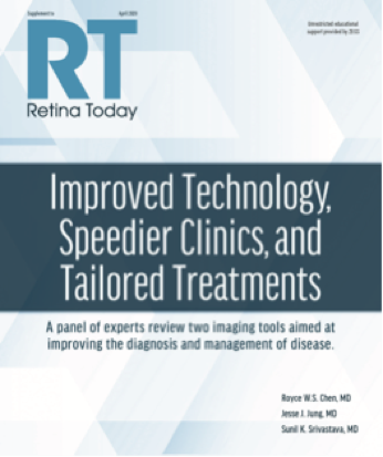
Improved Technology, Speedier Clinics, and Tailored Treatments
Imaging Geographic Atrophy in the Age of Treatment
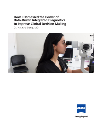
How I Harnessed the Power of Data-Driven Integrated Diagnostics to Improve Clinical Decision Making
Data Integration for Clinical Decision Making
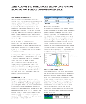
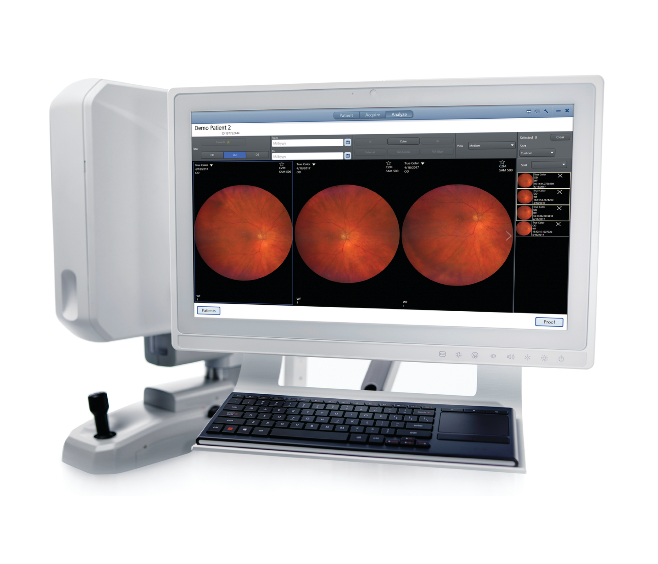
ZEISS CLARUS 500 Introduces Broad Line Fundus Imaging for Fundus Autofluorescence
About ZEISS
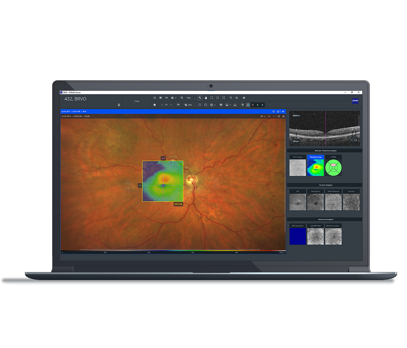
By combining premier imaging and at-a-glance history with interactive analysis, ZEISS Retina solutions support both your routine and complex patient management decision processes with an efficient, worry-free and cost effective solution.
Visit us online: zeiss.com/meditec/en/workflows/retina-workflow-idi-retina.html?utm_campaign=&utm_medium=eyetube&utm_source=bmc-eyetube-ret-workflow-2024
Follow ZEISS
en-INT_31_174_0286I
The statements of the doctors reflect only their personal opinions and experiences and do not necessarily reflect the opinion of any institution with whom they are affiliated. The doctors alone are responsible for the content of their experience reported and any potential resulting infringements. Carl Zeiss Meditec, Inc and its affiliates do not have clinical evidence supporting the opinions and statements of the doctors nor accept any responsibility or liability of the authors’ content.
Not all products, uses, treatment options and protocols referenced are officially approved in every market or supported by a product’s intended use in every market. Approved labeling and instructions may vary from one country to another. For country-specific product information, see the appropriate country website. Product specifications are subject to change in design and scope of delivery as a result of ongoing technical development.


%20(1)_1706629881.jpg?auto=compress,format&w=180)
%20(1)_1706630133.jpg?auto=compress,format&w=180)
%20(1)_1706630466.jpg?auto=compress,format&w=180)