


Search
Search
Show Description +
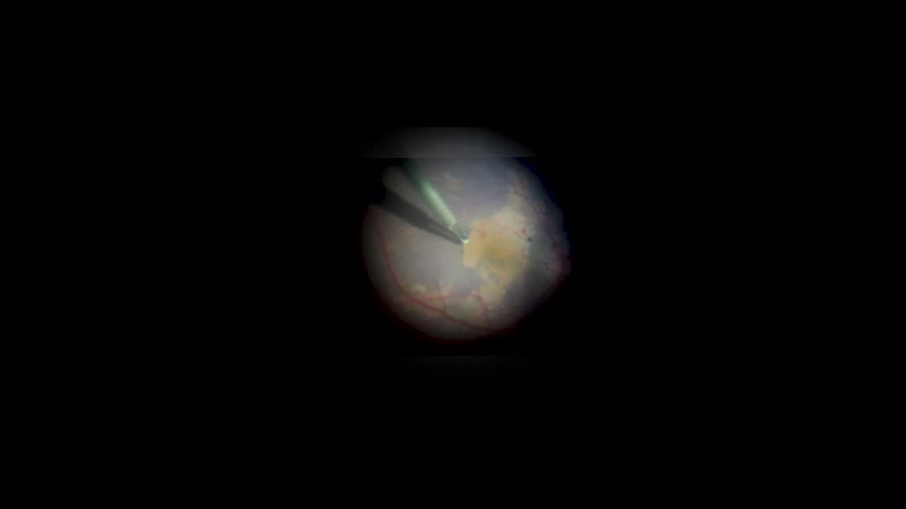
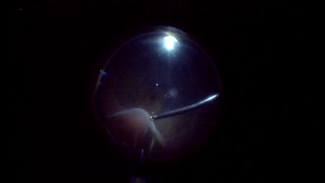
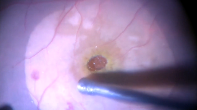
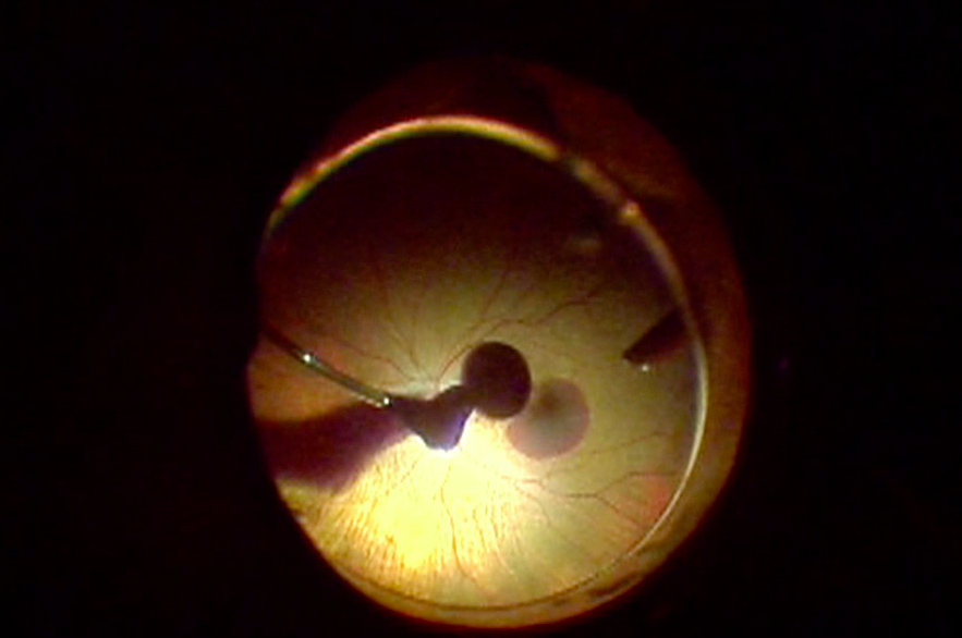
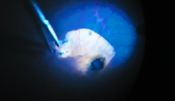
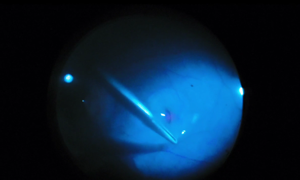
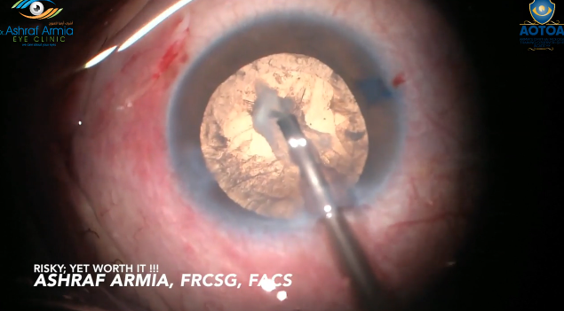
Mayank Bansal, MD, describes three cases of challenging intraocular foreign body (IOFB) removal. In the first case, the IOFB was stuck to the optic disc. Dr. Bansal performs removal of the IOFB without damaging the optic nerve or central retinal vessels. The second case had a retinal detachment with a 14-mm long IOFB. In the third case, Dr. Bansal shows the use of a 25G vitrectomy cutter to dissect the IOFB from the fibrous capsule, choroid, and sclera.
Posted: 8/12/2024
Mayank Bansal, MD, describes three cases of challenging intraocular foreign body (IOFB) removal. In the first case, the IOFB was stuck to the optic disc. Dr. Bansal performs removal of the IOFB without damaging the optic nerve or central retinal vessels. The second case had a retinal detachment with a 14-mm long IOFB. In the third case, Dr. Bansal shows the use of a 25G vitrectomy cutter to dissect the IOFB from the fibrous capsule, choroid, and sclera.
Posted: 8/12/2024
Sponsored Content
Sponsored Content





Please log in to leave a comment.