Using Intraoperative OCT to View the Canal During Hydrus Implantation

Show Description +

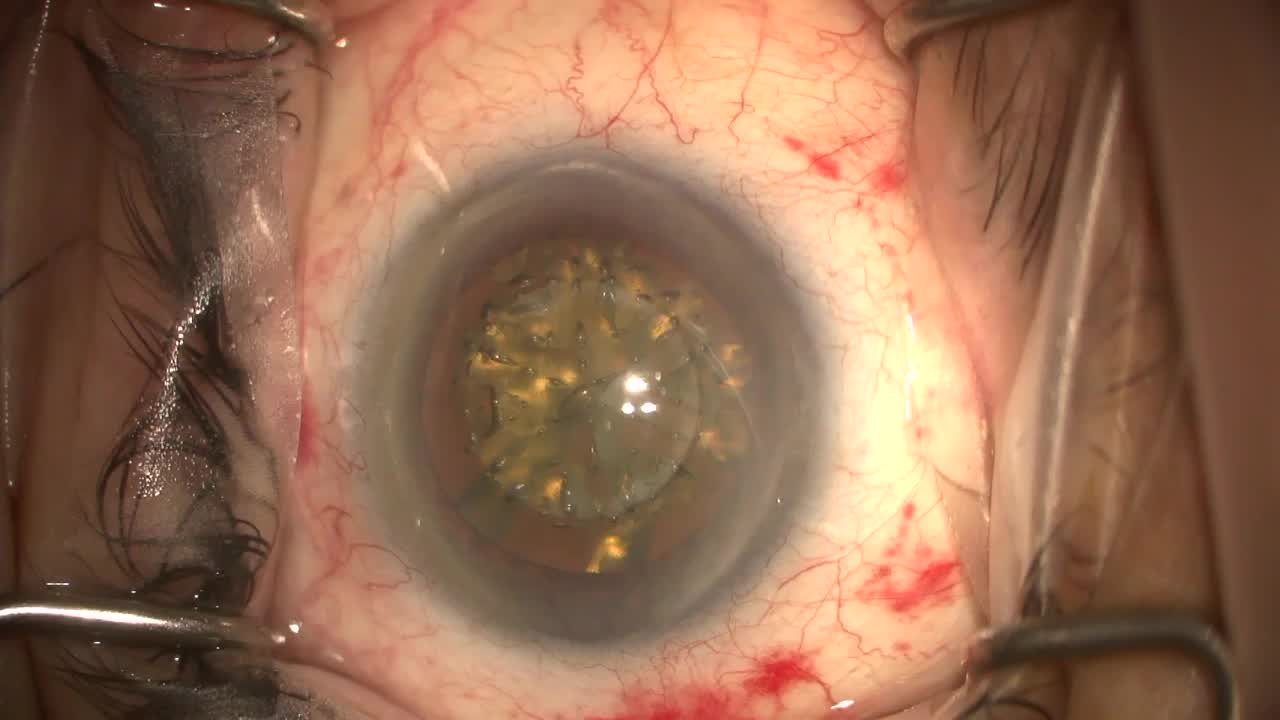
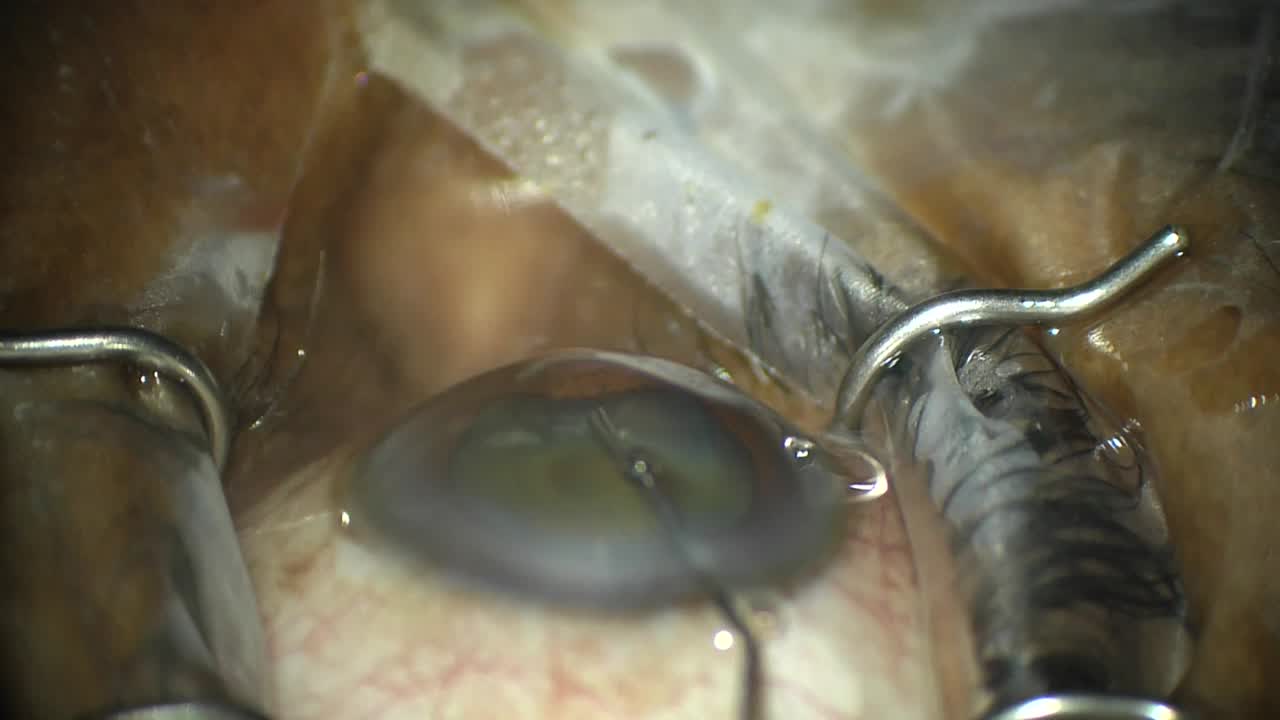
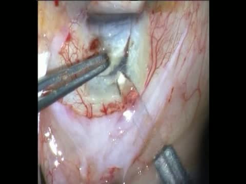
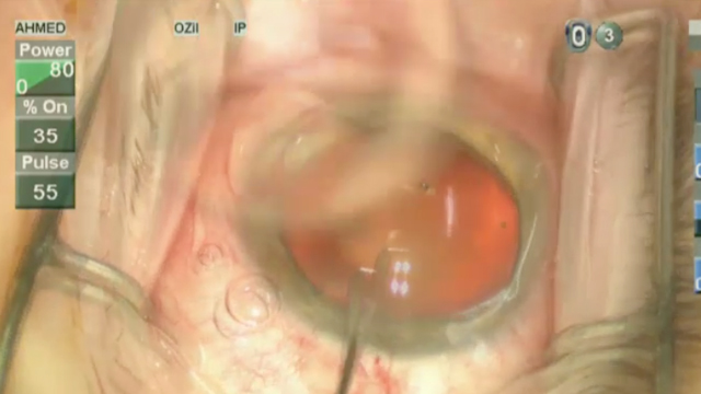
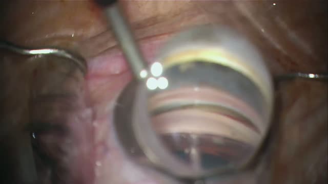
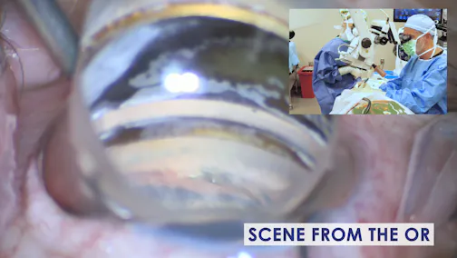
In this case of Hydrus Microstent (Ivantis) implantation, the intraoperative OCT demonstrates a large patent canal where the Hydrus is located but, beyond the Hydrus, the canal is more collapsed.

Posted: 11/30/2021

Using Intraoperative OCT to View the Canal During Hydrus Implantation

In this case of Hydrus Microstent (Ivantis) implantation, the intraoperative OCT demonstrates a large patent canal where the Hydrus is located but, beyond the Hydrus, the canal is more collapsed.

Posted: 11/30/2021

Please log in to leave a comment.

More in Glaucoma

Challenging Cases: From PRK to IOP

Radhika Rampat, MBBS, BSc(Hons), FRCOphth, and Marguerite B. McDonald, MD, FACS

Refractive Considerations in a Glaucomatous Eye

John P. Berdahl, MD; Natalia Morales, MD; and Justin Schweitzer, OD, FAAO

MIGS Unplugged LIVE 2026: A Candid Conversation on IG

Iqbal Ike K. Ahmed, MD, FRCSC; Arsham Sheybani, MD; Sahar Bedrood, MD, PhD; and I. Paul Singh, MD